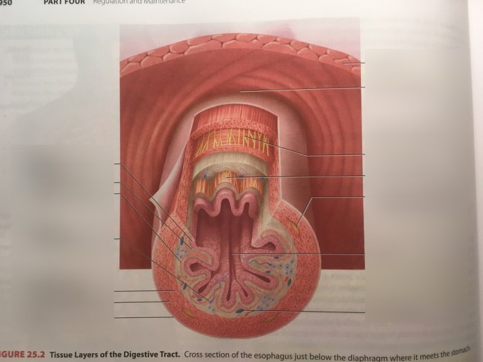
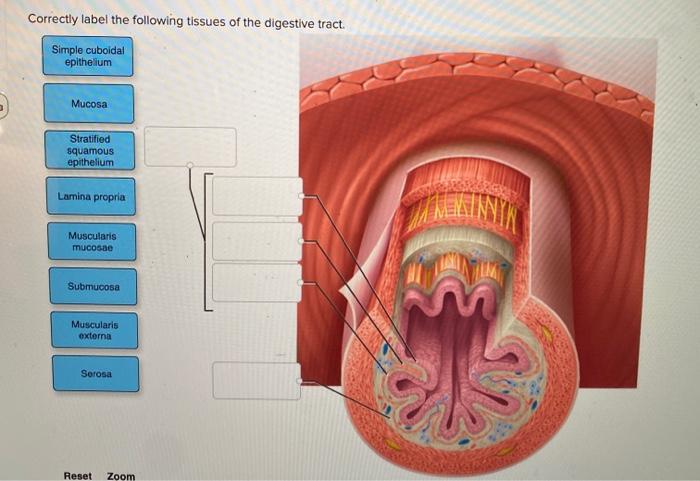
Correctly label the following tissues of the digestive tract. – Correctly labeling the tissues of the digestive tract is crucial for accurate diagnosis and effective treatment. Mislabeling can lead to incorrect diagnoses, inappropriate therapies, and adverse patient outcomes. This article provides a comprehensive guide to the histological features, immunohistochemical markers, and molecular techniques used to correctly label digestive tract tissues.
Introduction
Correctly labeling tissues of the digestive tract is crucial for accurate diagnosis and treatment of gastrointestinal diseases. Mislabeling can lead to incorrect diagnoses, inappropriate treatments, and potentially severe consequences for patients.
Methods for Labeling Tissues
Histology
Histology involves examining tissue samples under a microscope after they have been stained with dyes. It provides information about the tissue’s structure and cellular composition.
Immunohistochemistry, Correctly label the following tissues of the digestive tract.
Immunohistochemistry uses antibodies to detect specific proteins within tissues. It allows for the identification and differentiation of different cell types based on their protein expression.
Molecular Techniques
Molecular techniques, such as in situ hybridization and DNA sequencing, analyze the genetic material within tissues. They provide information about gene expression and mutations, which can aid in diagnosing genetic disorders.
Histological Features of Digestive Tract Tissues

Esophagus
The esophagus is lined with stratified squamous epithelium, which transitions to columnar epithelium at the gastroesophageal junction.
Stomach
The stomach has a mucosal layer consisting of gastric pits, glands, and parietal cells. The muscularis layer is responsible for churning and mixing food.
Small Intestine
The small intestine is characterized by villi and microvilli, which increase the surface area for absorption. The mucosal layer contains goblet cells that secrete mucus.
Large Intestine
The large intestine has a mucosal layer with crypts and goblet cells. The muscularis layer is thicker than in the small intestine.
Rectum
The rectum has a similar structure to the large intestine, but its muscularis layer is even thicker to facilitate defecation.
Immunohistochemical Markers for Digestive Tract Tissues: Correctly Label The Following Tissues Of The Digestive Tract.

| Tissue | Markers |
|---|---|
| Esophagus | Cytokeratin, p63 |
| Stomach | Gastrin, pepsin |
| Small Intestine | Villin, chromogranin A |
| Large Intestine | Mucin-2, CDX2 |
| Rectum | Cytokeratin 20, CD3 |
Molecular Techniques for Labeling Tissues

In Situ Hybridization
In situ hybridization uses labeled DNA or RNA probes to detect specific gene sequences within tissues. It provides information about gene expression patterns.
DNA Sequencing
DNA sequencing determines the order of nucleotides in a DNA molecule. It can identify mutations, genetic variants, and chromosomal abnormalities.
Case Studies
Mislabeling of digestive tract tissues can have serious consequences. For example, a case study reported a patient who was misdiagnosed with gastric cancer due to mislabeling of normal gastric mucosa as cancerous tissue.
FAQ Insights
What are the consequences of mislabeling digestive tract tissues?
Mislabeling can lead to incorrect diagnoses, inappropriate therapies, and adverse patient outcomes.
What are the different methods used to label digestive tract tissues?
Histology, immunohistochemistry, and molecular techniques are commonly used to label digestive tract tissues.
What are the advantages of using immunohistochemical markers to label digestive tract tissues?
Immunohistochemical markers can identify and differentiate different cell types in the digestive tract, providing valuable information for diagnosis and research.